Home

picnic pope squat normal fetal doppler ultrasound forgiven Mustache sandwich

Normal pulsed and color Doppler of fetal aortic artery (37 WG) | Download  Scientific Diagram
Normal pulsed and color Doppler of fetal aortic artery (37 WG) | Download Scientific Diagram

PDF] Fetal Doppler ultrasound assessment of ductus venosus in a 20 – 40  weeks gestation normal fetus in the Pakistani population | Semantic Scholar
PDF] Fetal Doppler ultrasound assessment of ductus venosus in a 20 – 40 weeks gestation normal fetus in the Pakistani population | Semantic Scholar

Umbilical arterial Doppler assessment | Radiology Reference Article |  Radiopaedia.org
Umbilical arterial Doppler assessment | Radiology Reference Article | Radiopaedia.org

Indications for Doppler ultrasound at 32 weeks gestation | Vinmec

Growth and Doppler ultrasound – Maternal Fetal Medicine

A Radiologist's Guide to the Performance and Interpretation of Obstetric  Doppler US | RadioGraphics
A Radiologist's Guide to the Performance and Interpretation of Obstetric Doppler US | RadioGraphics

The normal umbilical artery Doppler waveform is characterized by... |  Download Scientific Diagram
The normal umbilical artery Doppler waveform is characterized by... | Download Scientific Diagram

Peak Systolic Blood Flow in the MCA

Starship - Antenatal Doppler Studies in High Risk Pregnancies

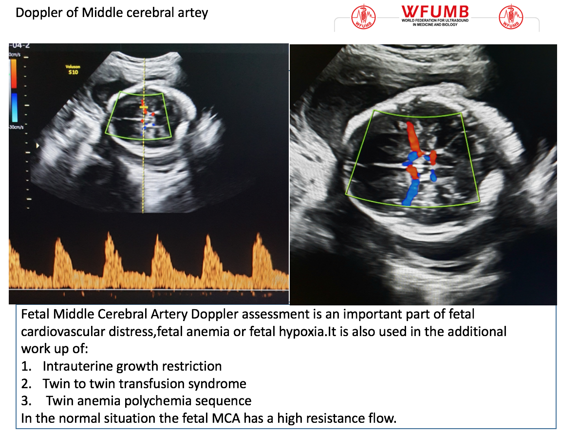
Ultrasound the Best #10: Doppler of the Fetal Middle of the Cerebral Artery  – WFUMB
Ultrasound the Best #10: Doppler of the Fetal Middle of the Cerebral Artery – WFUMB

Obstetric Dopplers – ULTRASOUNDPAEDIA

Fetal Middle Cerebral Artery Doppler Ultrasound Normal Vs Abnormal Image  Appearances | MCA USG - YouTube
Fetal Middle Cerebral Artery Doppler Ultrasound Normal Vs Abnormal Image Appearances | MCA USG - YouTube
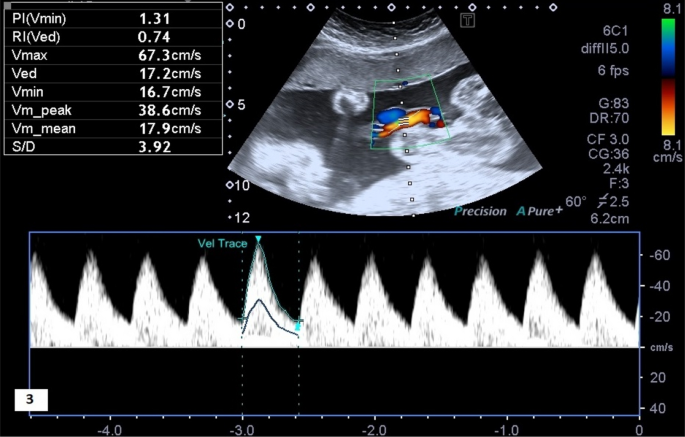
Normal umbilical artery doppler values in 18–22 week old fetuses with  single umbilical artery | Scientific Reports
Normal umbilical artery doppler values in 18–22 week old fetuses with single umbilical artery | Scientific Reports

Normal Doppler imaging of MCA and UA at 18 weeks of gestational age in... |  Download Scientific Diagram
Normal Doppler imaging of MCA and UA at 18 weeks of gestational age in... | Download Scientific Diagram

Assessment of Fetal Compromise by Doppler Ultrasound Investigation of the  Fetal Circulation | Circulation
Assessment of Fetal Compromise by Doppler Ultrasound Investigation of the Fetal Circulation | Circulation

Umbilical Artery Doppler Assessment: A How-To Guide | Empowered Women's  Health
Umbilical Artery Doppler Assessment: A How-To Guide | Empowered Women's Health

Assessment of Fetal Compromise by Doppler Ultrasound Investigation of the  Fetal Circulation | Circulation
Assessment of Fetal Compromise by Doppler Ultrasound Investigation of the Fetal Circulation | Circulation

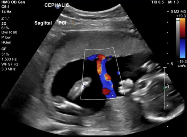
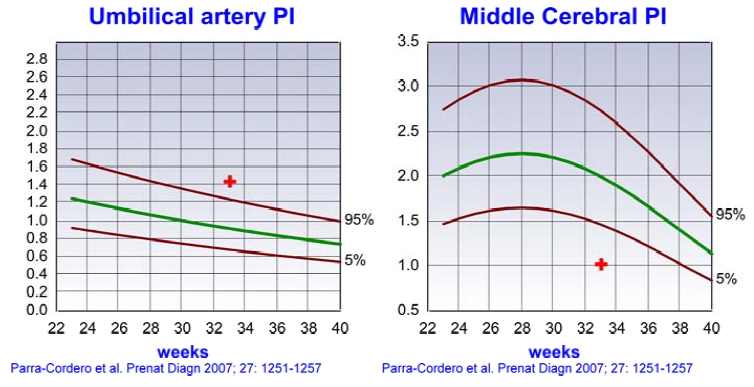
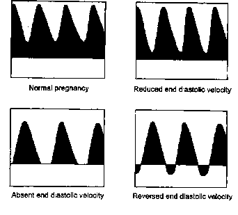
Doppler in Obstetrics

Ultrasound Examination 28-34 Weeks and Doppler - [Venus Med]

Umbilical Artery Doppler – Sonographic Tendencies

Umbilical Artery Doppler Assessment: A How-To Guide | Empowered Women's  Health
Umbilical Artery Doppler Assessment: A How-To Guide | Empowered Women's Health

A Radiologist's Guide to the Performance and Interpretation of Obstetric  Doppler US | RadioGraphics
A Radiologist's Guide to the Performance and Interpretation of Obstetric Doppler US | RadioGraphics

Doppler indices of the umbilical and fetal middle cerebral artery at 18-40  weeks of normal gestation: A pilot study. | Semantic Scholar
Doppler indices of the umbilical and fetal middle cerebral artery at 18-40 weeks of normal gestation: A pilot study. | Semantic Scholar
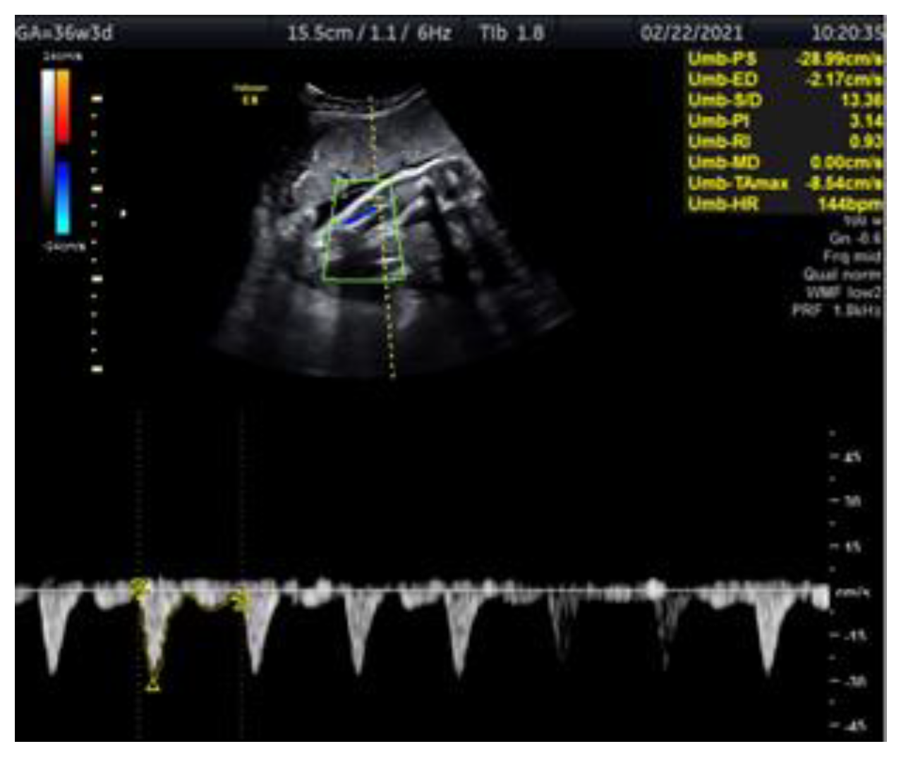
Medicina | Free Full-Text | Doppler Ultrasonography of the Fetal Tibial  Artery in High-Risk Pregnancy and Its Value in Predicting and Monitoring  Fetal Hypoxia in IUGR Fetuses
Medicina | Free Full-Text | Doppler Ultrasonography of the Fetal Tibial Artery in High-Risk Pregnancy and Its Value in Predicting and Monitoring Fetal Hypoxia in IUGR Fetuses
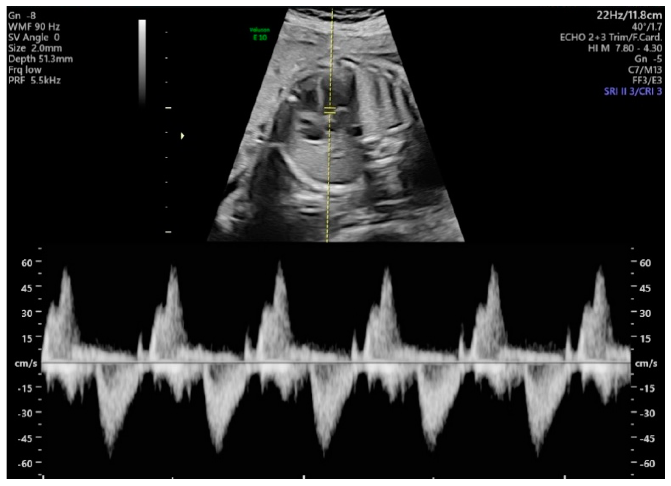
Diagnostics | Free Full-Text | Evaluation of the Fetal Left Ventricular  Myocardial Performance Index (MPI) by Using an Automated Measurement of  Doppler Signals in Normal Pregnancies
Diagnostics | Free Full-Text | Evaluation of the Fetal Left Ventricular Myocardial Performance Index (MPI) by Using an Automated Measurement of Doppler Signals in Normal Pregnancies